Geographical Distribution - World Wide
Habitat:- Entomoeba histolytica was discovered by Losch in 1875 in dysentery feces of a patient in Russia. It is found in the intestine and liver.
Marphology :- Entomoeba histolytica was discovered by Loch in 1875 in dysentery faces of a patient in Russia. It is found in the intestine and liver.
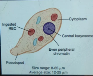
- Trophozoit Stage
- Pre-Cyst Stage
- Cyst Stage
1. Trophozoit Stage :-
- Trophozoit stage and Vegetative stage parasite are growing or feeding stage.
- Their shape is not fixed due to change of position.
- Their size is 18 to 40mm
- Its cytoplasm appears in two parts. Ectoplasm and Endoplasm
- Its nucleus is of spherical shape and the size of the nucleus keeps on increasing and decreasing from 4 to 6 mm.
- In fresh preparation the nucleus is not visible due to rapid movement but when the motility is reduced.So a slight outline of the nucleus becomes visible inside the body of the parasite.
- Stained preparation in nucleus structure in karyosome and nuclear membrane lilin network between karyosome and nuclear membrane.
- The trophozoite stage of Entamoeba histolytica is seen in freshly passed dysenteric stool.
- The trophozoit divide into 8 hours by Binary Fission.
Pre-Cyst Stage:-
It keeps on deceasing in size from samall about 10 to 20mm . It is round and slightly ovoid in shape with a blunt peripheral like projection. Pre-Cyst Stage in endoplasm , RBC and other Ingested food parficles are free.
Cyst Stage:-
- The installment spherical shape is about 10 to 20 mm in size and is surrounded by a highly refractory membrane called the kit wall.
- The early stage of installment consists of a single nucleus and a mass of two other structures and 1 to 4 chromate and chromidial bursae which are of scissor shape.
- Contains chromatin, RNA, DNA and phosphates.
- In mature cyst, glycogen mass and chromatoid bodies disappear.
- Mitotic division twice in the nucleus produces 1 to 2 to 4 nuclei.
- It clears cytoplasm.
Lifecycle:-
Entamoeba histolytica passes its lifecycle into only one host cell. There are 3 stages in its lifecycle.
- Trophozoit stage
- Pre-Cyst Stage
- Cyst Stage
Mature quadrinucleate is the infective form of which parasite.
When How Much Is Ingested By A Susceptible Person Through Contaminated Food And Water So it does its further development inside that person gut.
Fully developed installment enters Elementary canal and reaches Stomach.
Installment wall is resistant to gastric juice but digested by trypsin found in the intestine.
Exitation is obtained when the installment reaches the lower part of the valeum in the corpus.
Through this process, the cytoplasmic body moves beyond the wall and the installment divides into a single amoeba.In which there are 4 nuclei and 4 to 8 amoebulae.
This young amoebulae are activaly motile and get buried in the tissue.In spherically large, they keep it in the sub-mucous layer of the intestine as a normal habitat.
It Grow On This And Multiply By Binary Fission. The trophozoite stage of the parasite is responsible for producing amoebiasis lesion.
This parasite resides in the intestinal speech and causes acute dysentery due to which a large number of trophozoites are discharged along with the faces.
Pathogenicity of entamoeba Histolytica:-
The incubation period of Entamoeba histolytica in humans is 4 to 5 days.
Clinical Features:-
The term amoebiasis refers to those conditions that are clinically which is produced by invading different areas of the human host of Entamoeba histolytica.
Laboratory Diagnosis:-
The definitive diagnosis of amoebiasis depends on the demonstration of Entamoeba histolytica.Where is Entamoeba histolytica getting discharged from? Culture cannot be done in routine diagnosis.
Immunological test is not helpful in the diagnosis of intestinal infection but is helpful for diagnosis in extra intestinal amoebiasis.
Intestinal Amoebiasis :-
These are two types.
- Acute Amoebic Dysentery
- Chronic Amoebic Dysentery
Acute Amoebic Dysentery:-
In this, the stool sample is collected directly in a container of wide mouth and it is not diluted during examination.
First of all look at the stool with naked eyes. The stool is usually semi-fluid brownish black in color. And fecal material is found in some solsules and in blood and mucus. It is acetic in reaction
On microscopic examination, cellular exudates are found to be few. RBCs become clumped and appear redish yellow and yellowish green.
Culture and serological tests are not usually done in the routine. Serology is usually negative in early cases.
Chronic Amoebic Dysentery:-
Microscopic Examination of patients with chronic Amoebiasis in case of colon amoebic ulcer under direct microscope and Histopathology.


Comments
Post a Comment
Thanks to Come on Comment section